Cellular Profile of Wound Tissue
Under normal “uncompromised” circumstances skin wounds are “seen” to progress through
the stages of inflammation, new tissue formation and new tissue remodelling -
The cellular profile of wound tissue changes with time after injury.
Initially, inflammatory cells that are responsible for clearing wound site debris
(such as neutrophils and monocyte-
Consequently, the cellular profile of a wound can be used to describe its maturity. Wound tissues with a relatively high proportion of inflammatory cells are considered less mature (less advanced) than similar tissues containing relatively few inflammatory cells.
The relative contribution of inflammatory cells (neutrophils & monocyte-
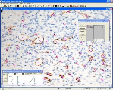
Cell specific histological staining techniques are used to label and measure the involvement of specific cellular lineages (see below).
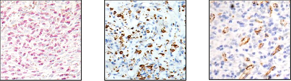
(Chloroacetate Esterase staining)
(ED-
(anti-
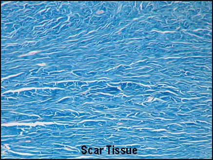
Scar Assessment
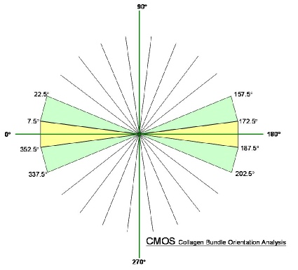
Scar Tissue Analysis is performed using our in house Cica Scar Analysis System (C-
C-
The output generated by C-
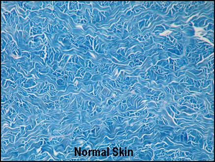
Scar tissue is composed of a collagen bundles
largely orientated parallel to the surface of the skin; whereas, normal skin has
a more multi-
Severe scars have more horizontally-
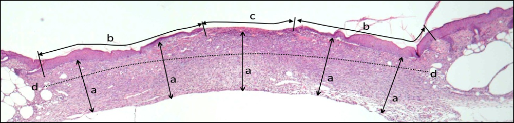
Routine H&E staining of a diabetic wound showing histological planimetry measurements:
a) Granulation tissue depth
b) Epithelial coverage
c) Open wound
d) Wound width measurement

 Back
Back