Home About Us Services Relevant Links Contact Us
wound contraction

Rate of Wound Closure


The impact of agents on wound closure is assessed using excisional wound models. Wound closure, which is measured by computer
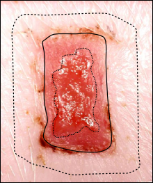
-assisted planimetry from calibrated images taken at intervals during the study, can be differentiated into its components:- Wound Re-epithelialisation & Wound Contraction (see below)


Original wound area

Area closed by contraction

Healing Wound

Area closed by re-epithelialisation

Open wound area remaining

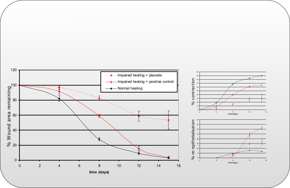
Data showing % wound area remaining (relative to the original wound area on day 0) ± s.e.m. for a diabetes-impaired wound healing model .

The components of wound closure (% wound re-epithelialisation and % wound contraction) are also displayed, demonstrating a reduced ability of wound closure by contraction in diabetes-impaired wounds accompanied by a slight increase in wound closure by re-epithelialisation.

Treatment with a growth factor combination therapy (positive control) demonstrates a positive modulation of both components .


Wound Planimetry


Diagram showing wound planimetry measurements in a normal wound healing model.


Rate of Wound Closure Wound Colour Profile Histological Assessments Tensiometry

Navigation


Back